
Cystic Adnexal Lesions
Mindy M. Horrow, MD, FACR, FSRU, FAIUM
Director of Body Imaging
Einstein Medical Center, Philadelphia, PA
Professor of Radiology
Jefferson Medical School
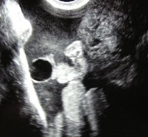
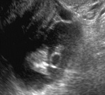
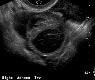
Ovum is surrounded by cumulusoophorus within the mature follicle
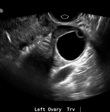
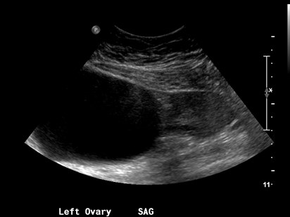
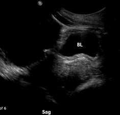
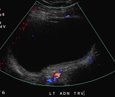
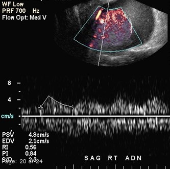
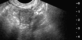
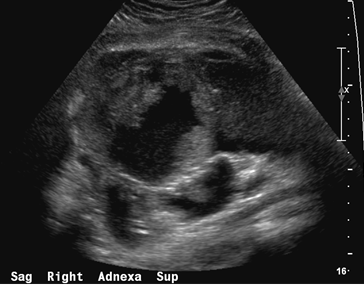
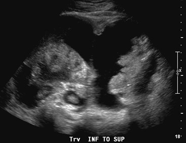
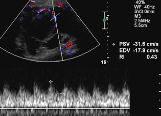
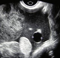
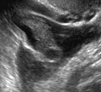
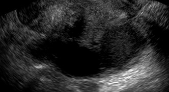
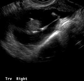
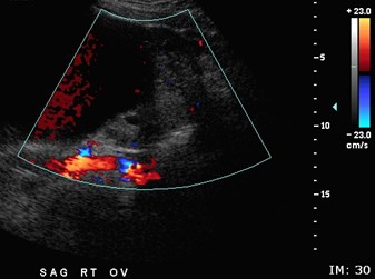
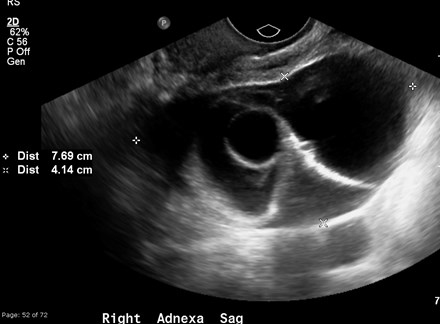
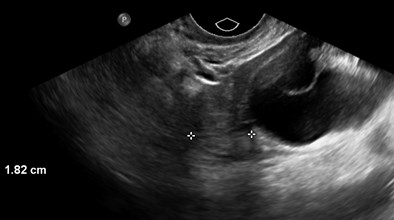
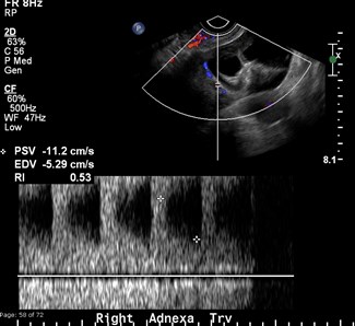
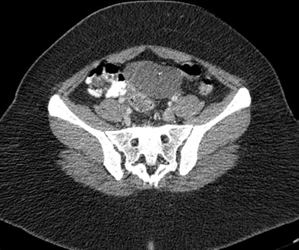
23 year old
Para-ovarian cyst
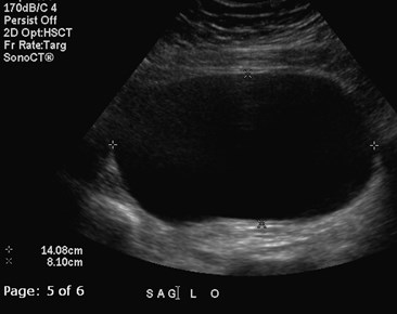
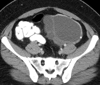
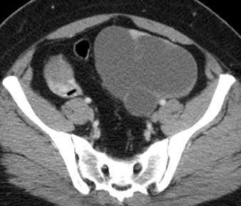
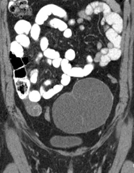
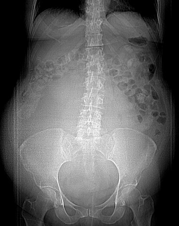
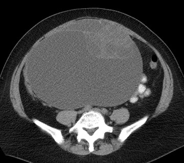
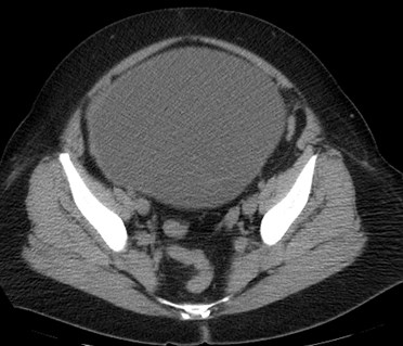
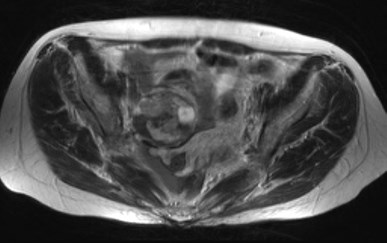
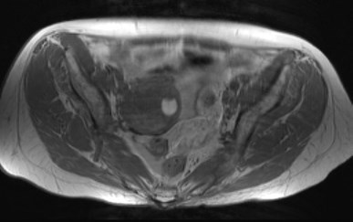
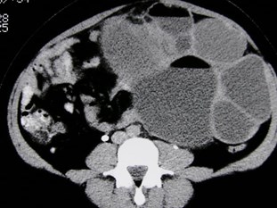
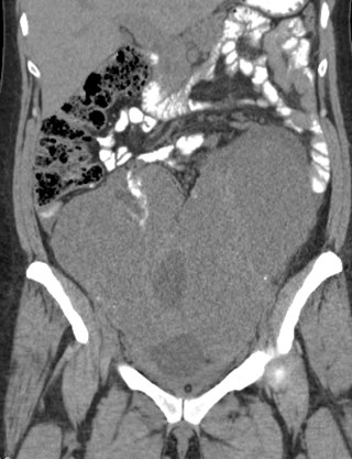
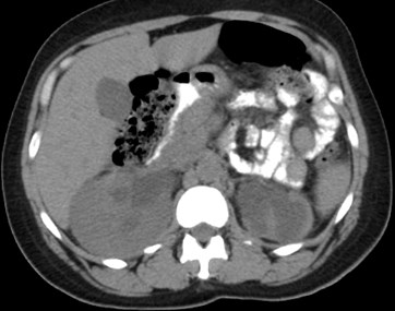
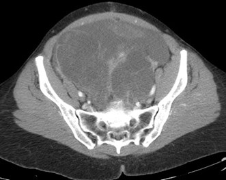
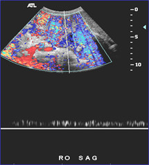
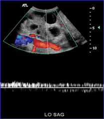
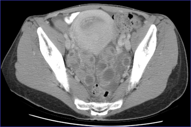
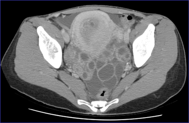
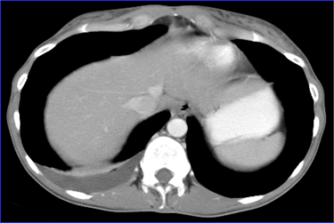
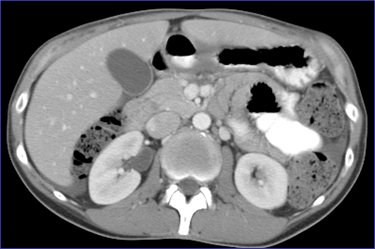
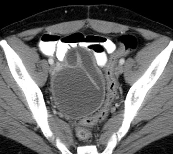
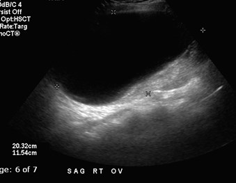
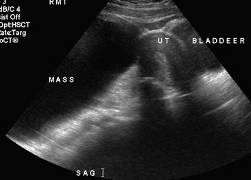
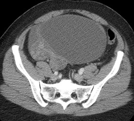
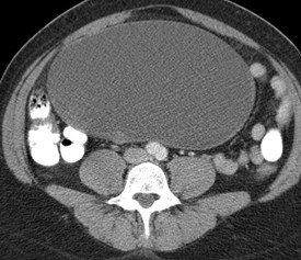
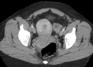
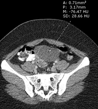
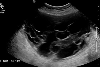
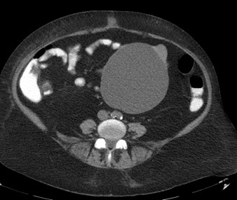
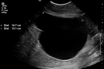
52 year old woman with abdominal distention
Mucinous Cystadenoma
Mucinous cystadenoma
•Almost always multilocular
•Septations often most easily appreciated on ultrasound
•May be extremely large
•Often contain fluid components with different densities orsignal
•80% of all mucinous ovarian neoplasms are smoothwalled benign cystadenomas
–10 – 15% are low grade malignancy
–5 – 10% are malignant
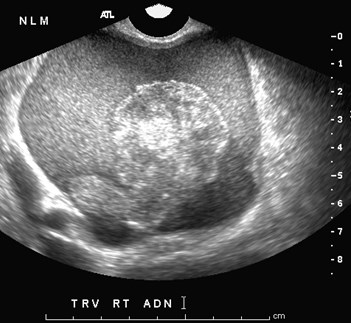
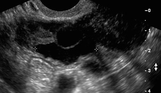
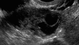
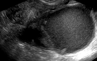
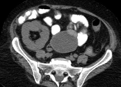
42 year old
What is the next step?
1.Follow up in 1-2 menstrual cycles
2.MR
3.PET
4.laparoscopy
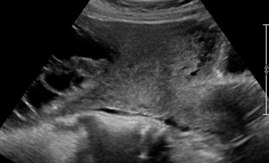
Serous cystadenoma
Serous cystadenoma
•Unilocular or multilocular cystic mass,homogeneous attenuation or signal, thin regularwall
•60% serous ovarian neoplasms are benign
–15% low malignant potential
–25% malignant
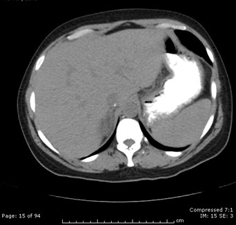
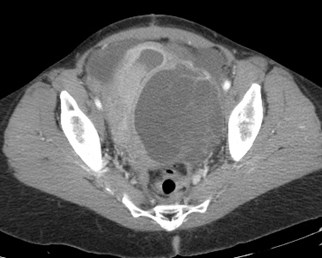
42 year old woman with distention
Borderline mucinous ovarian tumor

50 year old woman
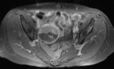
T1
T2
+C
Papillary Serous Tumorlow malignant potential
Borderline (low malignant potential)Neoplasms
•More proliferation of papillary projections
•More common in younger patients
•Much better prognosis than ovariancarcinomas
–Require staging
–Surgery may be ovarian/uterine sparing
–Adjuvant chemotherapy less likely
46 year old woman
Features of dermoid AND mucinous ovarian tumor
Collision Tumors
•Coexistence of 2 adjacent but histologicallydistinct tumors
•Ovarian collision tumors quite rare
•Most commonly composed of teratoma withcystadenoma or cystadenocarcinoma
•Consider when tumor has features that wouldnot be common to a single tumor
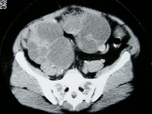
52 year old
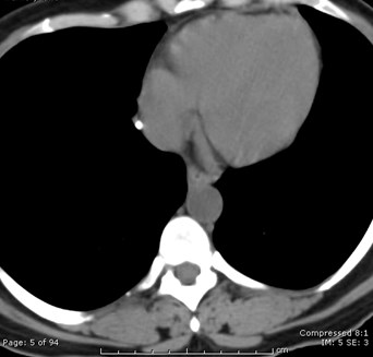
Serous cystadenocarcinoma ofovaries
Metastatic lymph nodes contain calcifications
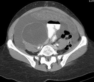
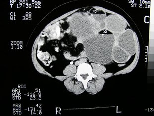
54 year old woman with colon cancer
Krukenberg Tumors, peritoneal carcinomatosis
Krukenberg Tumors
•Metastatic tumors to ovary containingmucin secreting “signet ring” cells
•Originate from GI tract, particularly colonand stomach
•Other common metastases to ovaries frombreast, lung, contralateral ovary
•Account for 10% of ovarian tumors duringreproductive years
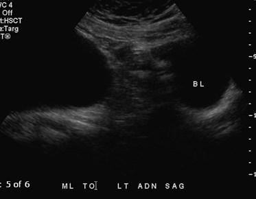
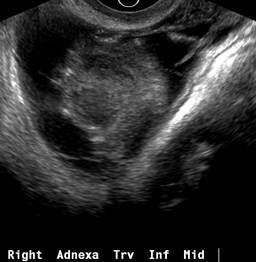
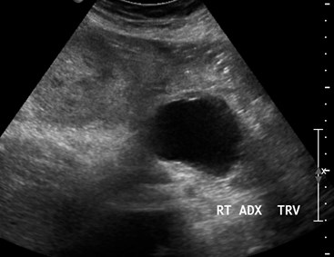
Diffuse pelvic pain
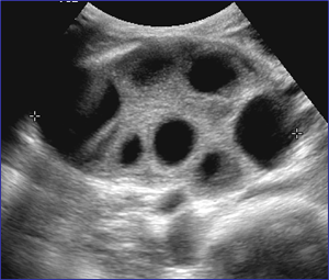
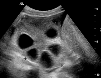
Ovarian HyperstimulationSyndrome
OHSS
•Potentially life threatening complication of ovulationinduction or stimulation
•Occurs during luteal phase of menstrual cycle or earlypregnancy
•Imaging hallmark: bilaterally symmetric enlarged ovariescontaining cysts in presence of ascites
•Shift of fluid acutely out of intravascular space resultingin ascites and hemoconcentration
–Most severe: hypercoaguable, renal and hepatic dysfunction,thromboembolic events, ARDS
Two different patients with same diagnosis
Hydatid of Morgagni (paratubal cyst)
10-06
If patients with underlying hydrosalpinx are re-infected,images may appear worse than clinical situation
9-07
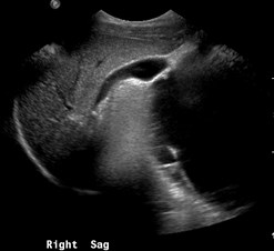
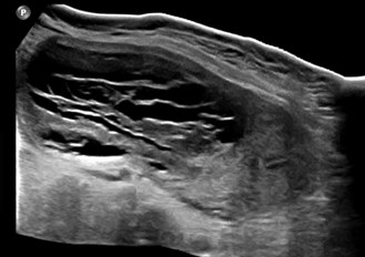
38 year old with chronic pelvic pain
Peritoneal Inclusion Cyst
Peritoneal Inclusion Cysts
•Type of pseudocyst in which fluid produced byovary is trapped within adhesions
•Usually result of endometriosis, PID, surgery
•Characteristic features
–No wall- irregular passive shape conforms to contoursof surrounding structures
–Ovary entrapped within
–Adhesions may become thickened and vascularized
Laing, Allison. Radiographics 2013;32:1621
3 non ovarian related “cysts”
•Paratubal (Morgagni) cysts
•Hydrosalpinx
•Peritoneal inclusion cysts
34 year old with 3 days of severe pelvic pain
Ovarian Torsion
17 year old with acuteright pelvic pain
Atypical Dermoid
49 year with distention
Cystic Degeneration of Myoma
64 year old woman with renal tx, CT for abdominal pain
4-2012
Follow up 3-2013